

Hone your POCUS interpretation and clinical reasoning by working through the interactive cases below.
Case 1: a surprising turn of heart
A 40-ish male, previously healthy on no regular meds, presents with 3 days of orthopnea and SOBOE. He denies pedal edema. Yesterday he had scant frothy pink sputum; no fever or chills. He has no VTE risk factors.
Review of systems is unremarkable. Past medical history is notable only for a reportedly normal kidney biopsy as a child for an episode of self-resolving hematuria caused by a “traumatic injury.”
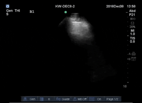
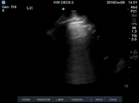
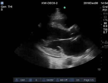
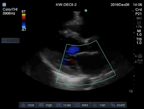
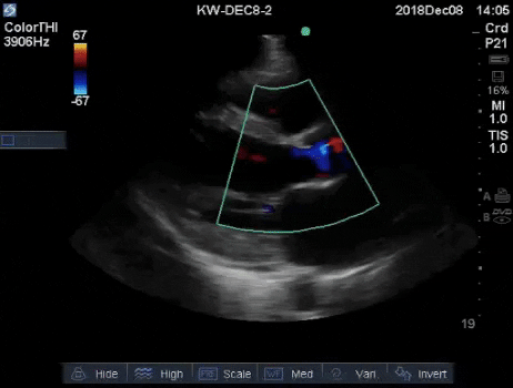
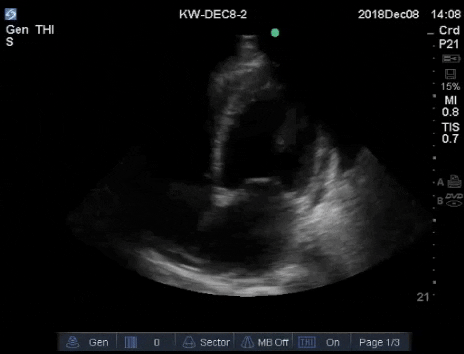
Below are representative images from the patient’s thoracic ultrasound.
Labs come back and reveal a Cr of 1100 (12mg/dL), K+ of 6.1, and a urinalysis showing 3+ Hgb and protein.
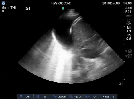
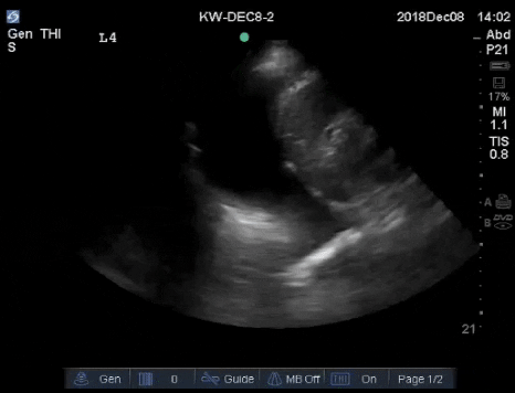
Here are his abdominal POCUS views.


Case Conclusion
The patient was admitted and a Cardiology-performed Echo confirmed the POCUS findings of a dilated cardiomyopathy with an EF of 25-35%. His renal failure was believed by the Nephrology team to be chronic, and likely a result of untreated IgA nephropathy as a child. He was initiated on dialysis. His extensive cardiac workup revealed no cause for his dilated cardiomyopathy, and it was therefore believed that he suffered from uremic cardiomyopathy as a consequence of prolonged markedly elevated urea levels.