

Hone your POCUS interpretation and clinical reasoning by working through the interactive case below.
Case #2: To lasix or not to lasix?
This was a 52 year old obese female who presented with severe anasarca and AKI (Cr 200-300 micromol/L). She has recently been diagnosed with biopsy-proven membranous nephropathy, and is awaiting a workup for secondary causes. There is no prior history of cardiac, renal, or liver disease. On admission, she was started on a lasix infusion given her anasarca, though she was on room air and BNP was normal at 70. She was diuresing well with the infusion, so 2 days earlier was stepped down to intermittent bolus lasix doses.
However, just prior to handover, the resident gets called because of increased oxygen requirements and progressive dyspnea. By the time the resident arrives at the bedside, the patient has been placed on BiPAP by the RT (previously on room air). There is a note of decreased urine output over the past 24 hours, as well as a mild non-productive cough. Vitals at the time were as follow: HR 90 (regular), BP 178/98, GCS 15.
The resident at the bedside suspects flash pulmonary edema and orders 180mg IV lasix and 5mg IV metolazone; however, there is no immediate response.
This cases poses the ubiquitous question: is this a case of TOO MUCH diuretic (no urine output because the patient has been dried out) or TOO LITTLE diuretic? Or is there something else going on entirely?
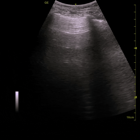
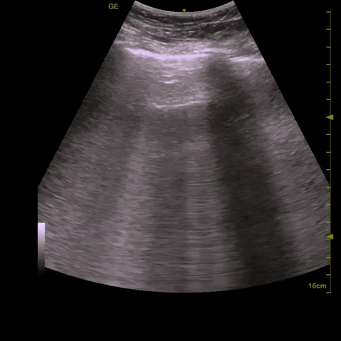
The POCUS team is on the scene. Here’s the first image , looking at her IVC.
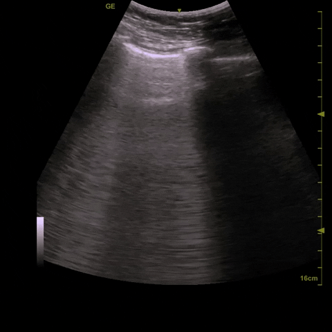
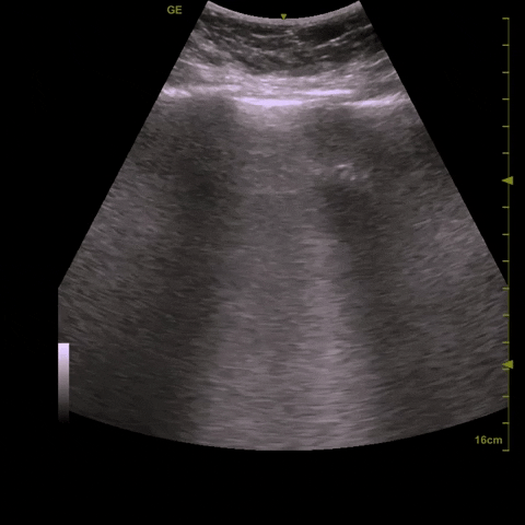
And here’s her lung study, starting with the left lung:
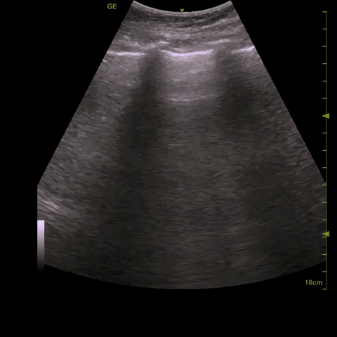
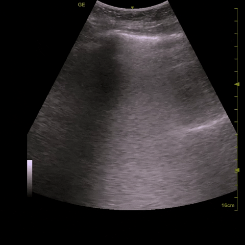
And the right:
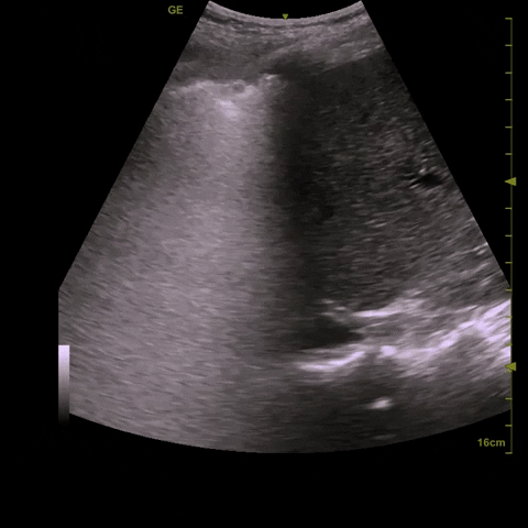
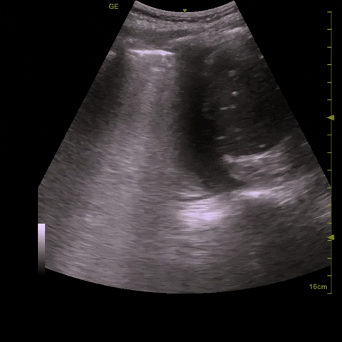
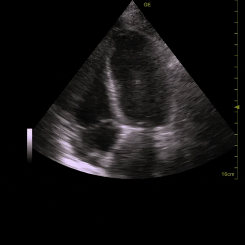
Now we’re getting into the cardiac scan. Here we’ve started at with the parasternal long-axis view:
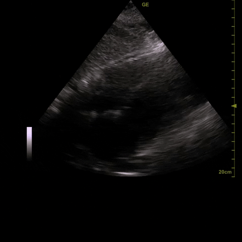
And now the apical and subxiphoid views:
Case conclusion
First, let’s summarize our image interpretation. The IVC is somewhat equivocal; it was performed without BiPAP (good), but in a patient with increased intra-abdominal pressure, making interpretation challenging. Remember, the IVC is going to be most valuable at extremes; in cases like this, it doesn’t add much to the clinical picture. The lungs, however, are very helpful here: the diffuse B-lines in a dependent gradient with largely smooth pleura, coupled with bilateral pleural effusions, are textbook for cardiogenic pulmonary edema. Finally, our cardiac scan reveals decreased LV function (which we can say despite slightly suboptimal images), a normal right ventricle (normal size, normal visual TAPSE and free wall excursion), and no pericardial effusion. Taken together, they all point to cardiogenic pulmonary edema as the cause of the patient’s respiratory decompensation.
Given that the patient had failed a hefty diuretic dose, the Nephrology service was consulted and the patient was started on IHD. She was significantly symptomatically improved within a few hours, and was back on room air by the next day. Labs showed a BNP of >2000, and a workup for the cause of her cardiomyopathy was initiated. A Cardiology-performed Echocardiogram suggested possibly Takotsubo’s cardiomyopathy, though the final diagnosis is still unclear.