
Hone your POCUS interpretation and clinical reasoning by working through the interactive case below.
Case #20: A Spooky Belly
TBD
Initial POCUS EXAM
Zone R3 (right axilla)

Abdominal Ultrasound - RUQ

Zone R4 (right lung base)

Abdominal Ultrasound - LUQ

Cardiac ultrasound
PLAX

PSAX

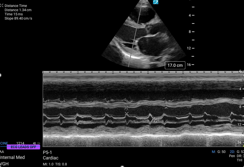
EPSS
A4C

Additional POCUS EXaMs
IVC - Long Axis

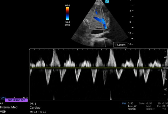
VExUS - Hepatic Vein
VExUS - Intrarenal Vein

IVC - Short Axis

VExUS - Portal Vein

Case Continued
A thoracentesis is performed at the bedside, clear straw coloured fluid is obtained and a total of 1.5L is drained. Fluid analysis as below:

Serum LDH 477
Serum Protein 70
Serum albumin 30
A paracentesis is performed at the bedside, somewhat cloudy orange fluid is obtained, and a total of 2.4L is drained. Fluid analysis as below:

Case Continued
The patient was diagnosed with congestive heart failure resulting in right sided pleural effusion and ascites from portal hypertension. They were treated with intravenous furosemide and after a period of days repeat images were obtained.
Post Diuresis - IVC Short Axis
Post-Diuresis - IVC Long Axis


Case Conclusion:
This case demonstrates the utility of bedside ultrasound for evaluation of pleural effusions and ascites, and in helping determine the etiology. Bedside ultrasound was further utilized to facilitate procedures for therapeutic benefit and for fluid analysis which supported the clinical diagnosis. Finally this case demonstrates the utility of bedside ultrasound in evaluation of decongestion following diuresis.