Hone your POCUS interpretation and clinical reasoning by working through the interactive case below.
Case #11: Double trouble
Written by Dayna Ingves, Adam D’Ovidio, and Farida Almarzooqi
Ms J is a 62 F with cirrhosis, ESRD on peritoneal dialysis, admitted on with PD peritonitis. She was treated with appropriate IV antibiotics. Her PD catheter was removed and she was started on HD.
Subsequently, she developed SOB, cough, and worsening hypoxemia (oxygen requirements increased from RA to 2L NP) that did not improve despite a higher fluid removal with UF. She also had persistent leukocytosis.
CXR at the time:
Pulmonary edema L > R. Moderate left pleural effusion. Bilateral lower lobe airspace opacification L > R.
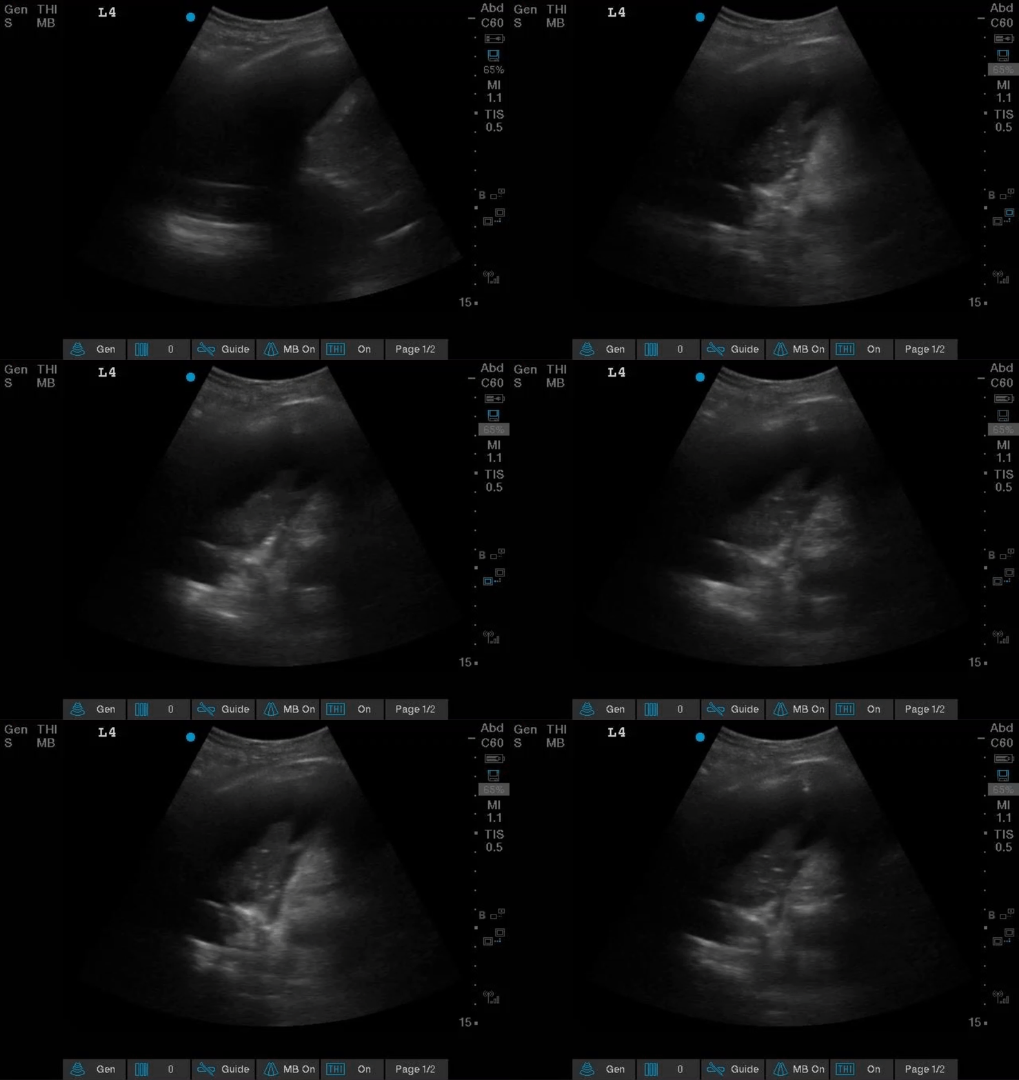
Differentials for her source of sepsis was intra-abdominal sepsis versus pneumonia. In addition, there was concern if her ascites/distended abdomen was causing diaphragmatic splinting and affecting her respiratory status. POCUS consulted to perform a diagnostic paracentesis, and a diagnostic + therapeutic thoracentesis.
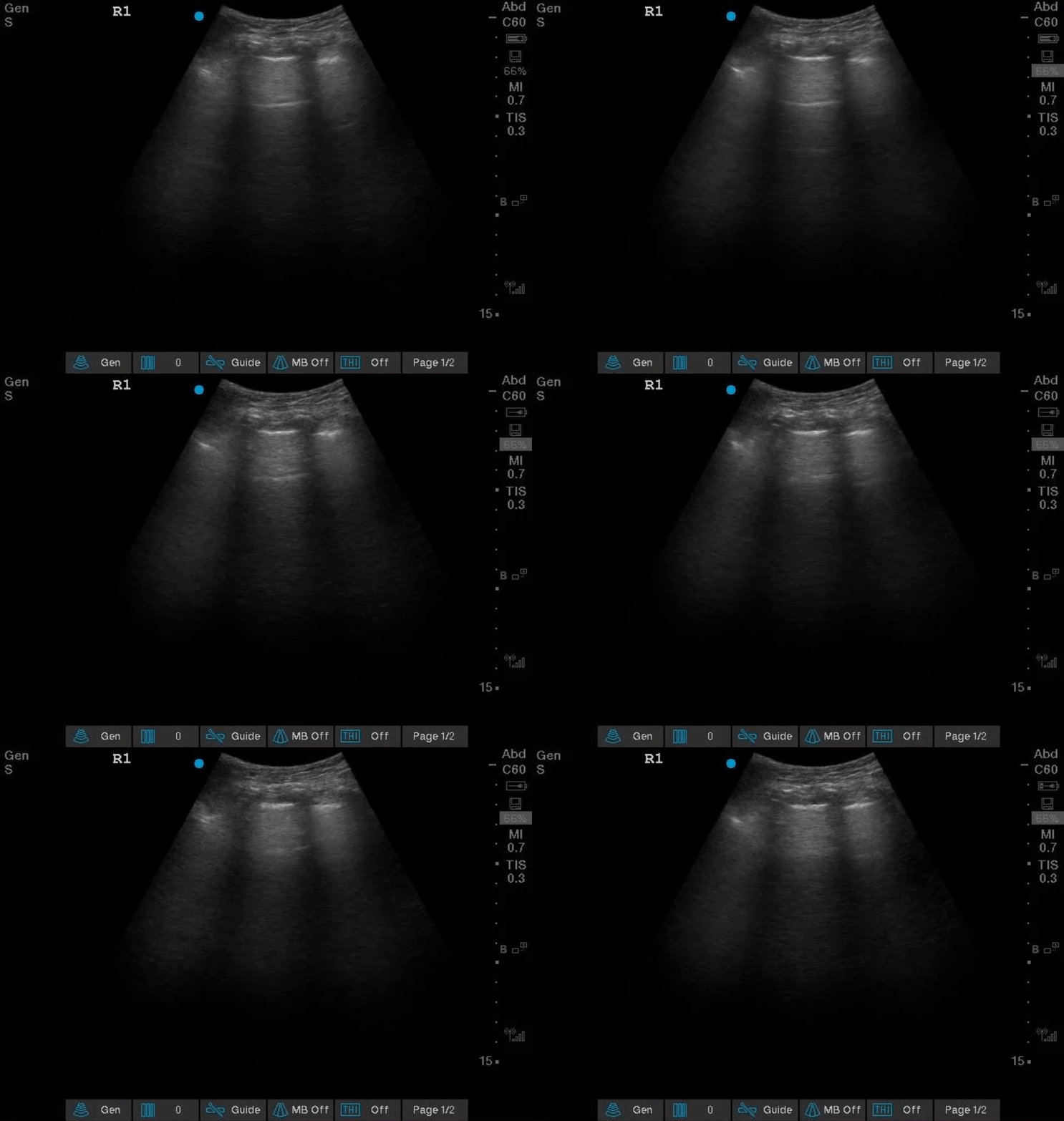
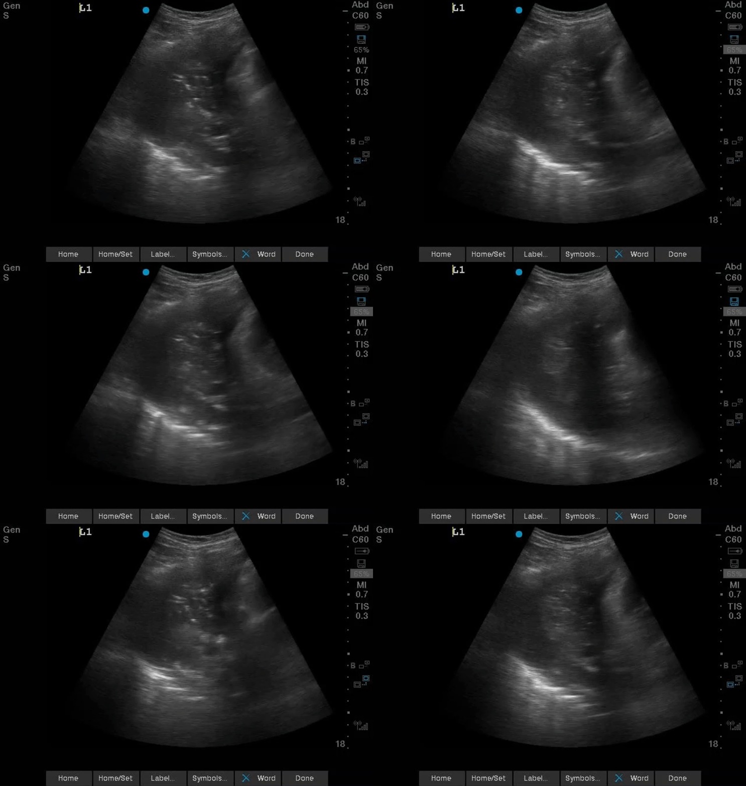
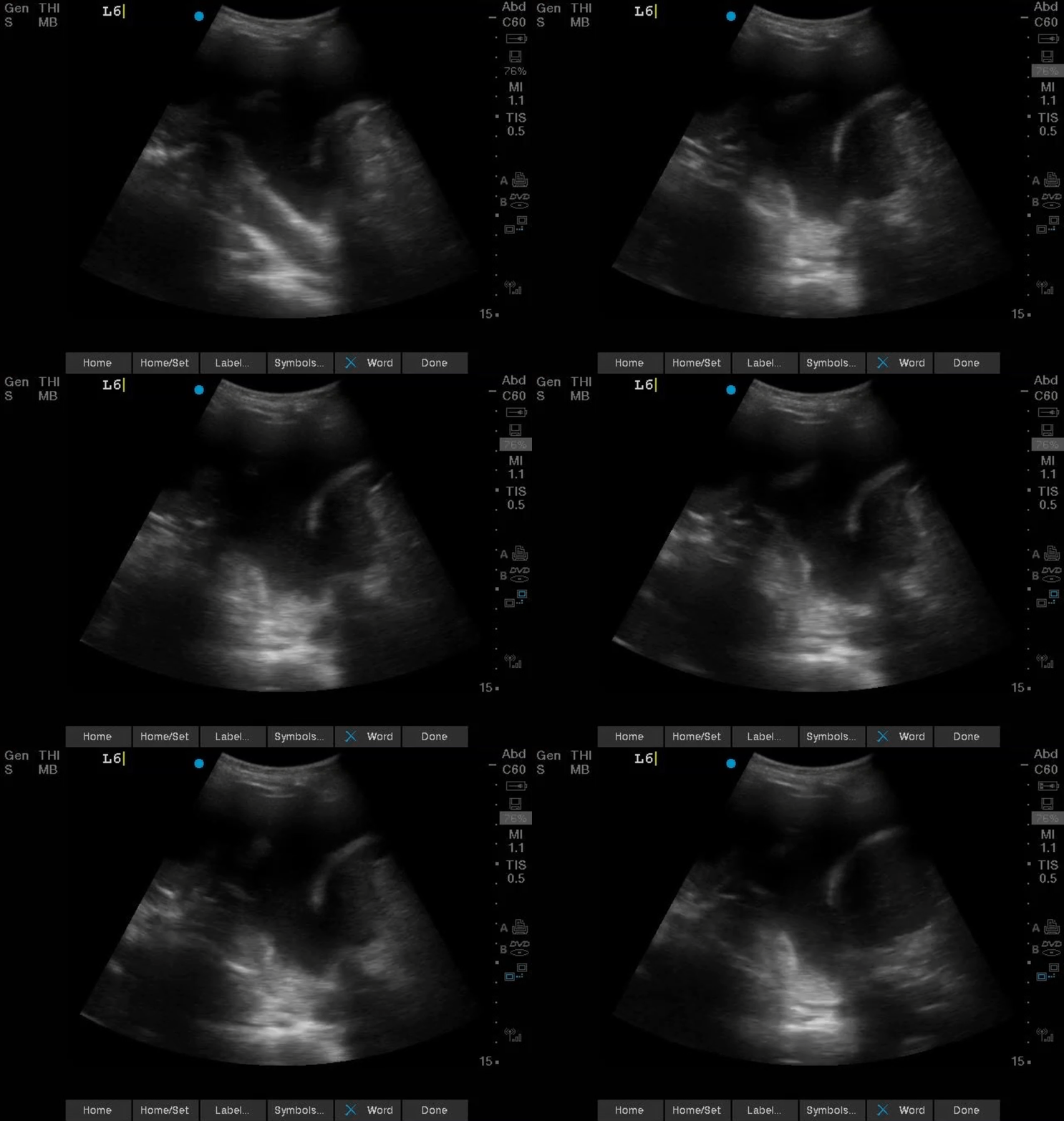
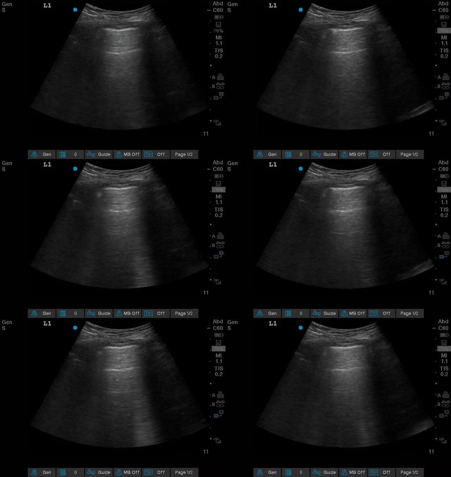
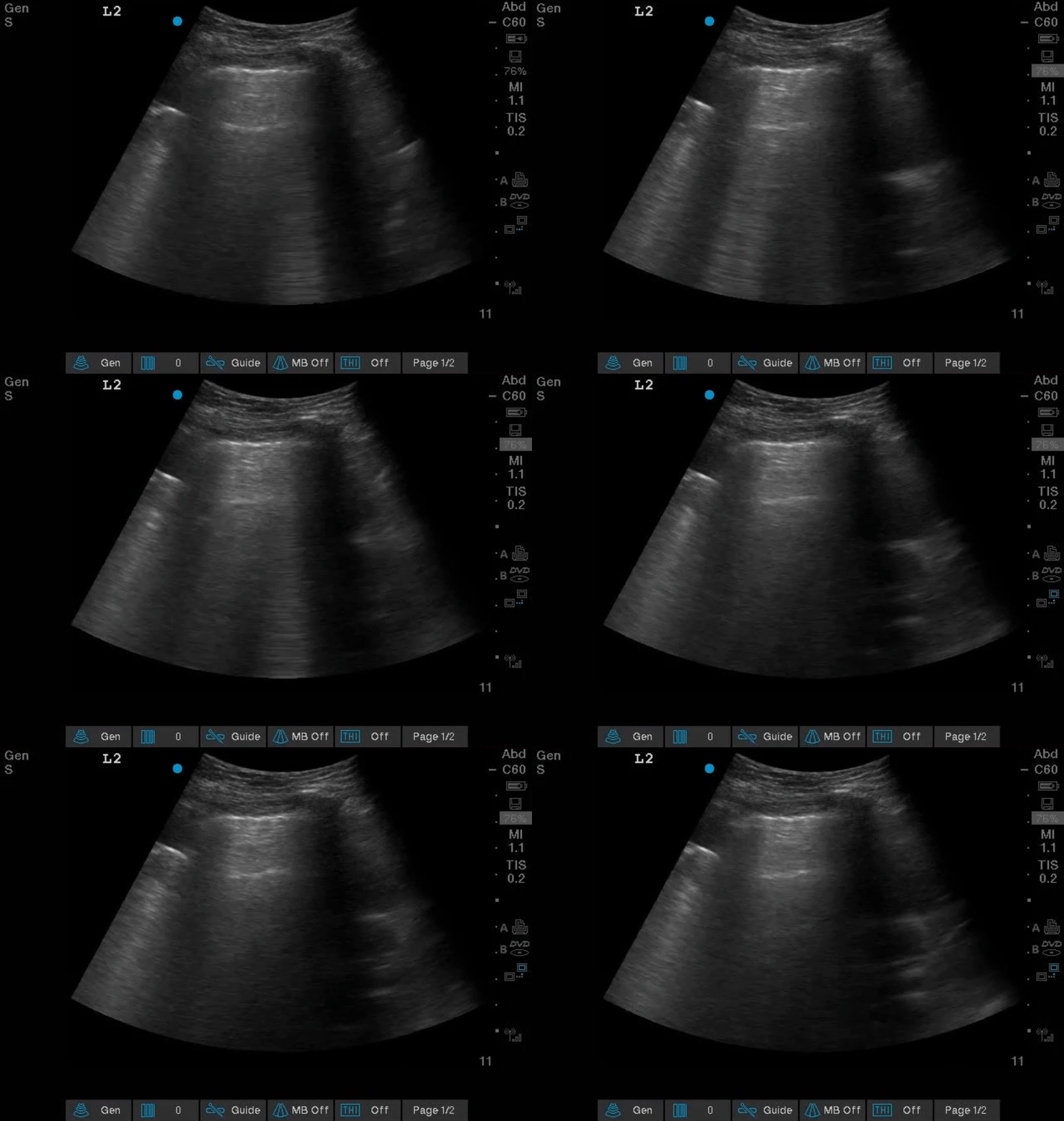
An initial lung POCUS exam was done.
RIGHT UPPER ANTERIOR LUNG FIELD (R1)
LEFT UPPER ANTERIOR LUNG FIELD (L1)
Air Bronchogram Basics
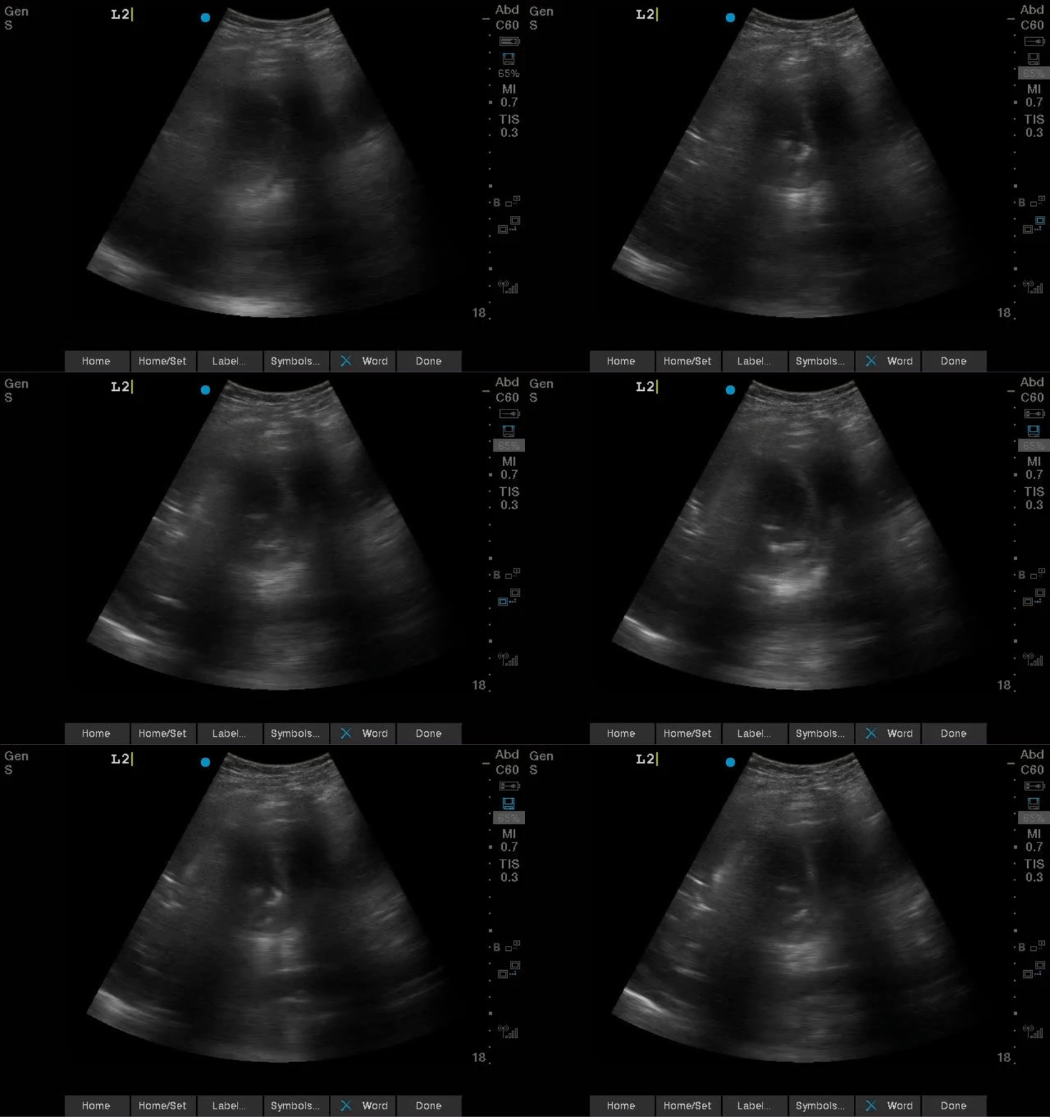
More images from the left lung…
Additional clinical information during our exam…
As patient was too frail to sit upright for us to access her effusion posteriorly, she was turned to the right lateral decubitus position.
During this maneuver, she started coughing++ continuously and unable to stop, and she desaturated significantly.
She was laid on her back again and positioned at 45 degrees which relieved the cough. However, she needed Optiflow 60% to maintain her saturation.
Thoracentesis was deferred.
Case Progression
With respiratory physiotherapy and suctioning, her mucus plug resolved with marked reduction in oxygen supplemental need.
The following is her repeated left thoracic scan.
Next Steps
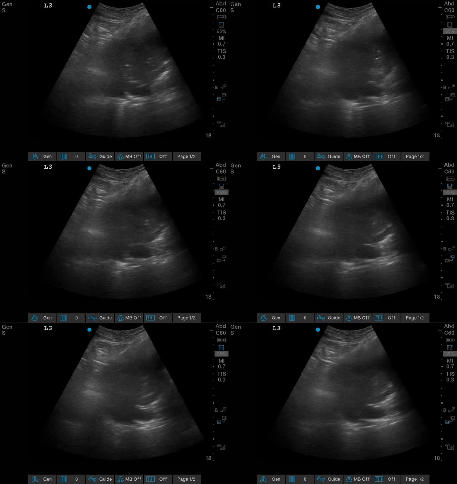
A diagnostic paracentesis was requested for source of leukocytosis and whether her PD peritonitis truly resolved. A POCUS exam of her RUQ is shown below:
Location of the largest pocket of ascites
As there was no other safe site for paracentesis due to the proximity of the bowel, this site was chosen just over the liver.
Can paracentesis be performed over the liver??
With the following safety considerations:
Can be done provided there is sufficient depth of fluid and there is no other safer access point
Use a small gauge needle (21 gauge)
Measure abdominal wall depth to confirm if 21G needle is long enough to reach the peritoneal space (remember that local anesthetic administration can increase soft tissue thickness)
Is there a risk of injuring the liver with the needle?
A cirrotic liver is >12kPa by fibroscan
For perspective: 1Pa is about the pressure of 1 sheet of paper. 1kPa is 1000 sheets of paper stacked on one another
It will be extremely difficult for a 21G needle to penetrate the liver! Liver biopsy is usually performed using a 16-18G needle.
POCUS Pearls
1.Size and location of pleural effusion relative to the underlying consolidated lung may indicate whether the atelectasis is due effusion compressive or not.
2.Type of air bronchograms (static vs. dynamic) may suggest a diagnosis for the consolidated lung and guide immediate interventions.
References:
1. Shah A, Oliva C, Stem C, Cummings EQ. Application of dynamic air bronchograms on lung ultrasound to diagnose pneumonia in undifferentiated respiratory distress. Respir Med Case Rep. 2022;39:101706. Published 2022 Jul 30. doi:10.1016/j.rmcr.2022.101706
2.Copetti, Roberto & Soldati, Gino & Copetti, Paolo. (2008). Chest sonography: A useful tool to differentiate acute cardiogenic pulmonary edema from acute respiratory distress syndrome. Cardiovascular ultrasound. 6. 16. 10.1186/1476-7120-6-16.
3. Eibenberger KL, Dock WI, Ammann ME, Dorffner R, Hörmann MF, Grabenwöger F. Quantification of pleural effusions: sonography versus radiography. (1994) Radiology. 191 (3): 681-4. doi:10.1148/radiology.191.3.8184046 - Pubmed
4. Balik M, Plasil P, Waldauf P, Pazout J, Fric M, Otahal M, Pachl J. Ultrasound estimation of volume of pleural fluid in mechanically ventilated patients. (2006) Intensive care medicine. 32 (2): 318. doi:10.1007/s00134-005-0024-2 - Pubmed
5. Lisi M, Cameli M, Mondillo S, et al. Incremental value of pocket-sized imaging device for bedside diagnosis of unilateral pleural effusions and ultrasound-guided thoracentesis. Interact Cardiovasc Thorac Surg. 2012;15(4):596–601. PMID: 22815326
6. Zanforlin A, Gavelli G, Oboldi D, Galletti S. Ultrasound-guided thoracenthesis: the V-point as a site for optimal drainage positioning. Eur Rev Med Pharmacol Sci. 2013;17(1):25–28. PMID: 23329520
